Three papers in the last few months, which feels like a decent enough excuse to do a quick roundup.
Micro-CT scanning tracks (Tash Prescott et al.)
First up is Tash Prescott with a paper just out in Fossil Studies:
How do you visualise what’s happening beneath the surface when a foot presses into sediment, without destroying your sample? Sounds straightforward. Isn’t really.
The standard approach is to physically section through track samples after formation. Works fine once, then your sample is gone. Tash and Ben Griffin used micro-CT scanning instead — non-destructive, full 3D volume. They made two separate track volumes using different substrate consistencies (‘soft’ and ‘very soft’) with alternating layers of sand and clay, then scanned them during indentation by a cadaveric pheasant foot.
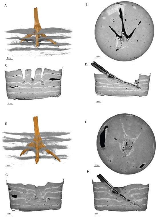
The CT reconstructions showed clear differences in sub-surface sediment displacement between the two consistencies, plus some complex features you’d never spot from surface morphology alone. Micro-CT gives us a way to look at volumetric track formation that doesn’t require splitting rocks or destroying experimental samples. Tracks are fundamentally three-dimensional structures, and most of what we’re interested in happens below the surface.
Penetrative track morphology (Ben Griffin et al.)
Ben Griffin, our post-doc, has been running discrete element method simulations looking at how tracks form in three dimensions — not just the surface impression but the whole subsurface volume.
The big finding: subsurface layers of a track made in soft sediment preserve foot motions (and morphology) much more reliably than the surface does. The surface gets distorted by sediment collapse, water drainage, all sorts of post-depositional processes. But look at layers just beneath the surface and you get a cleaner record of what the foot actually looked like when it pressed in. The subsurface layers in fact don’t change very much for a given foot and motion when you change substrate properties.
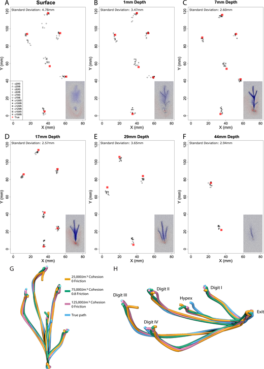
Ben systematically varied friction and cohesion using both a vertically indenting cylinder and a dynamic theropod-like tridactyl foot model. Higher friction produced taller, more extensive displacement rims throughout the track volume. Low cohesion made surface tracks deviate substantially from actual foot morphology — in extreme cases they looked deceptively like weathered tracks, which is worrying for anyone trying to interpret fossil preservation states.
Ben’s work supports motion reconstructions from penetrative tracks regardless of sediment conditions at formation, which is a strong result. The subsurface layers are surprisingly robust to changes in substrate properties — deeper than you’d think you can still trust what you’re seeing.
Digital range of motion analysis (Rebecca Lowes et al.)
Finally, Rebecca Lowes — a PhD student — has published her first paper in the Journal of Anatomy:
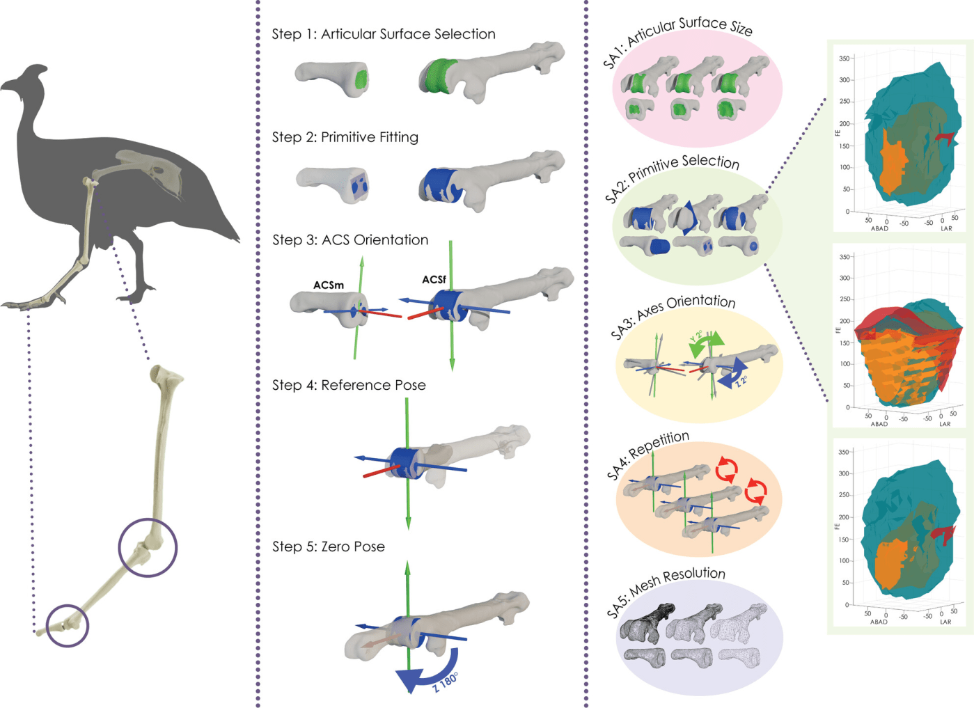
This one tackles something that sounds technical but is pretty fundamental to digital biomechanics. When you’re doing range of motion (ROM) analysis on 3D joint models — increasingly how we assess joint mobility in comparative anatomy — there are several subjective steps in building the model:
- How do you select the articular surfaces?
- Where do you place the joint centre of rotation?
- What reference pose do you start from?
Nobody had done a proper sensitivity analysis on this before. Rebecca ran one on a complete six-degree-of-freedom automated ROM workflow, testing how variation at each stage affects the final output. She used guineafowl ankle and tarsometatarsophalangeal joints.

The results show that ROM outputs are quite sensitive to variation in reference pose assembly, especially changes in articular surface selection and the primitive used to define the joint centre. This doesn’t necessarily mean individual studies are wrong — particularly when using maximum viable rotations to constrain biomechanical models — but it does make comparing results between studies and taxa much more problematic than we’ve realised.
Her recommendation (which I think is spot on) is that future ROM studies should provide complete joint models as supplemental data so other people can actually replicate the work. It’s a call for better transparency in a field where reproducibility matters.
What’s next?
All three tie into the broader EVOTRACK project — understanding how animal motion is preserved in the fossil record, from initial foot-sediment interaction through to reconstructing joint mobility and locomotor behaviour from the remains.
Well done to Tash, Ben, and Rebecca on these.

Leave a comment